
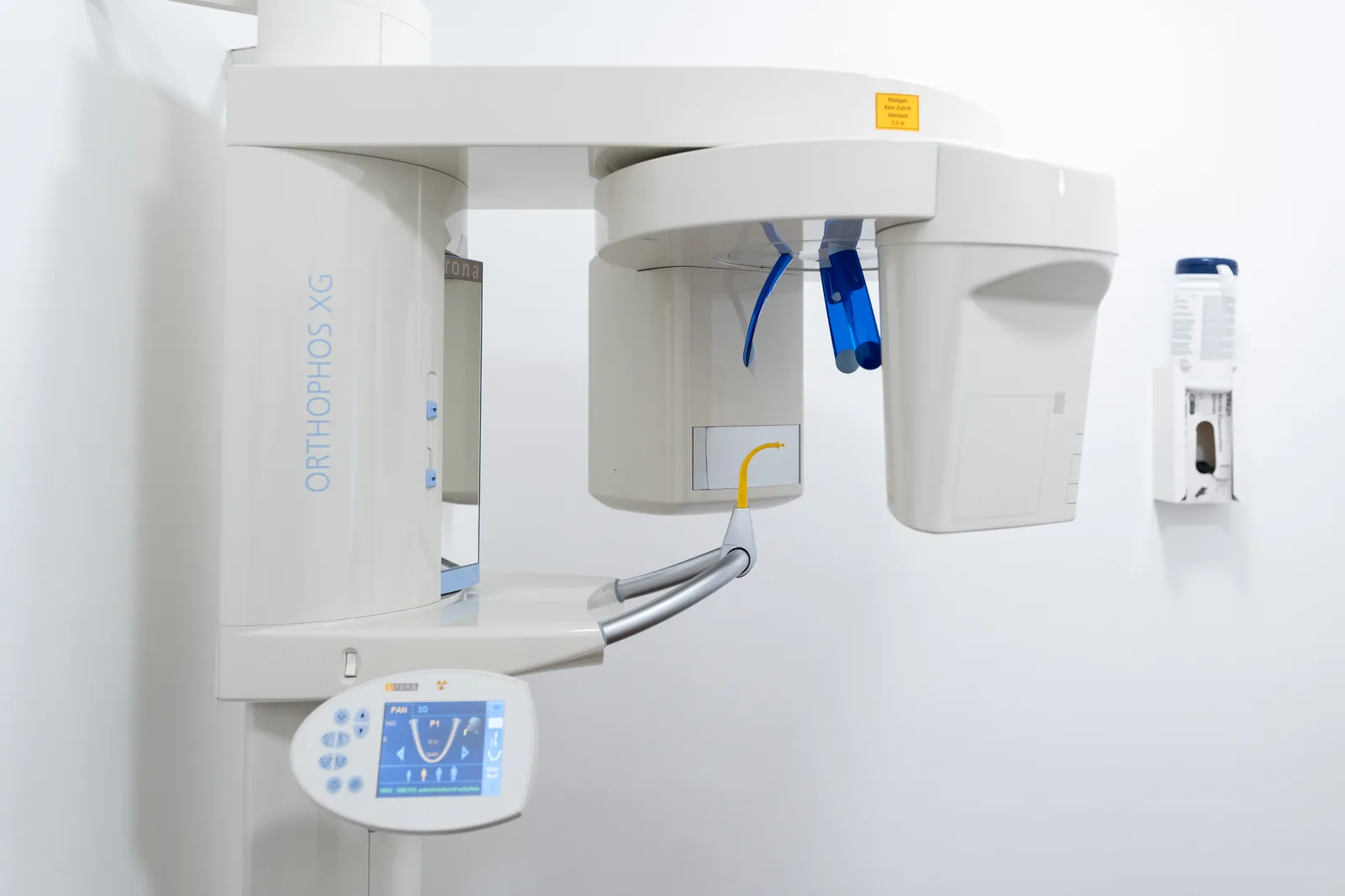
Often, for example, it is only through 3D imaging that it becomes possible to identify the exact width of the jawbone, or to assess the spatial course of an important nerve pathway and protect it accordingly.
The advantages of 3D X-ray (cone-beam CT):
- We can build a more accurate picture of the anatomical conditions, as the body's structures are also shown in depth. The images are also especially detailed, true to scale and free of distortion.
- In general, treatments such as implant placement can be planned more precisely, and can therefore usually be carried out more safely and gently.
- Compared with the commonly used computed tomography (CT), cone-beam CT significantly reduces radiation exposure.
How cone-beam CT works:
- 3D X-ray (cone-beam CT) produces individual cross-sectional images of the jaw or the whole head.
- These individual images can either be used directly for precise, to-scale measurements, or combined using computer software into a complete image of the jaw.
- The complete image is then available on screen, for example for computer-assisted 3D implant planning.
Video
Briefly explained – from our practice
The videos provide general information and do not replace individual advice; the course and outcome of treatment depend on personal circumstances. All videos have subtitles. More in our media library and on Instagram.
